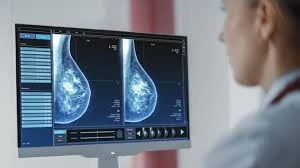
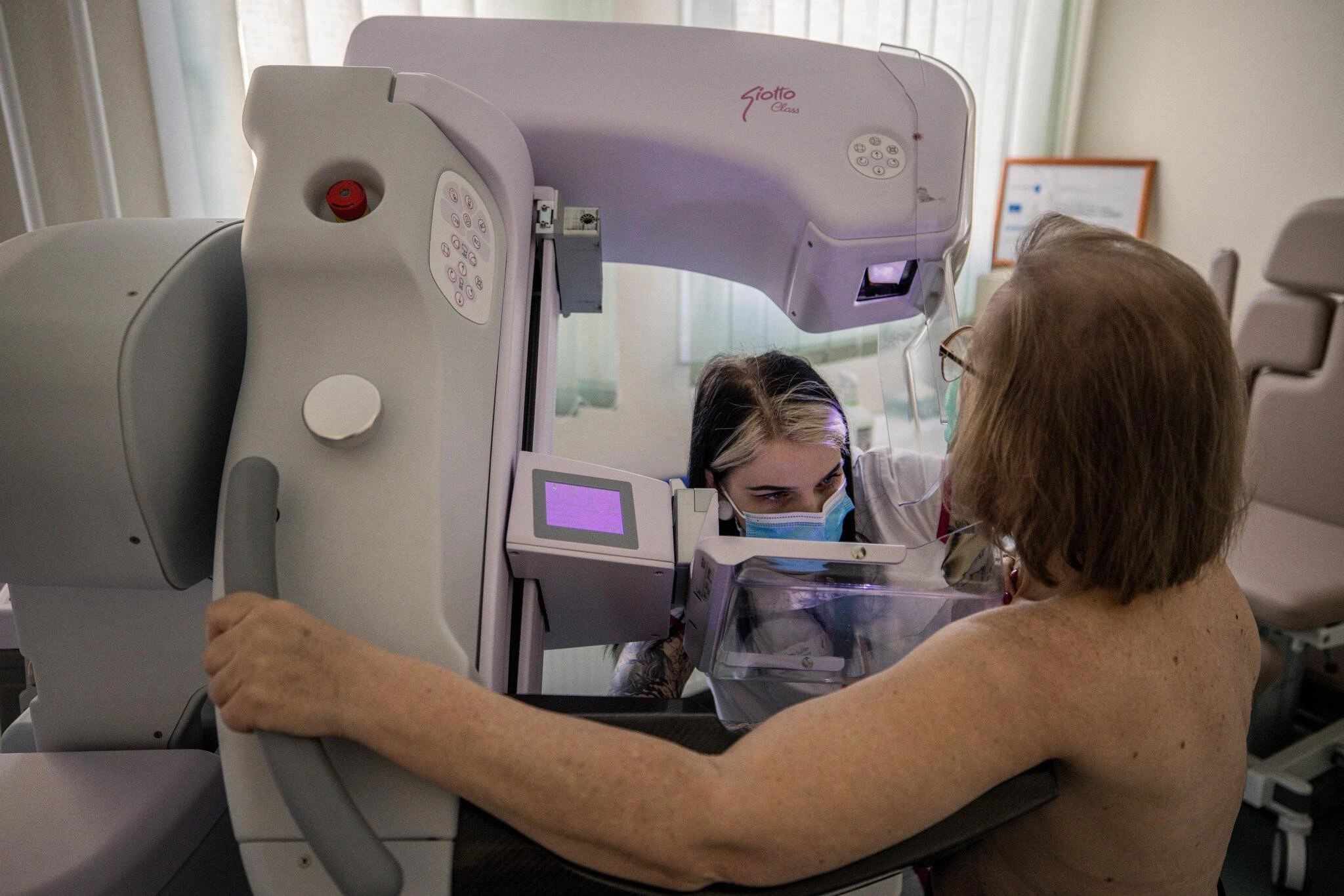
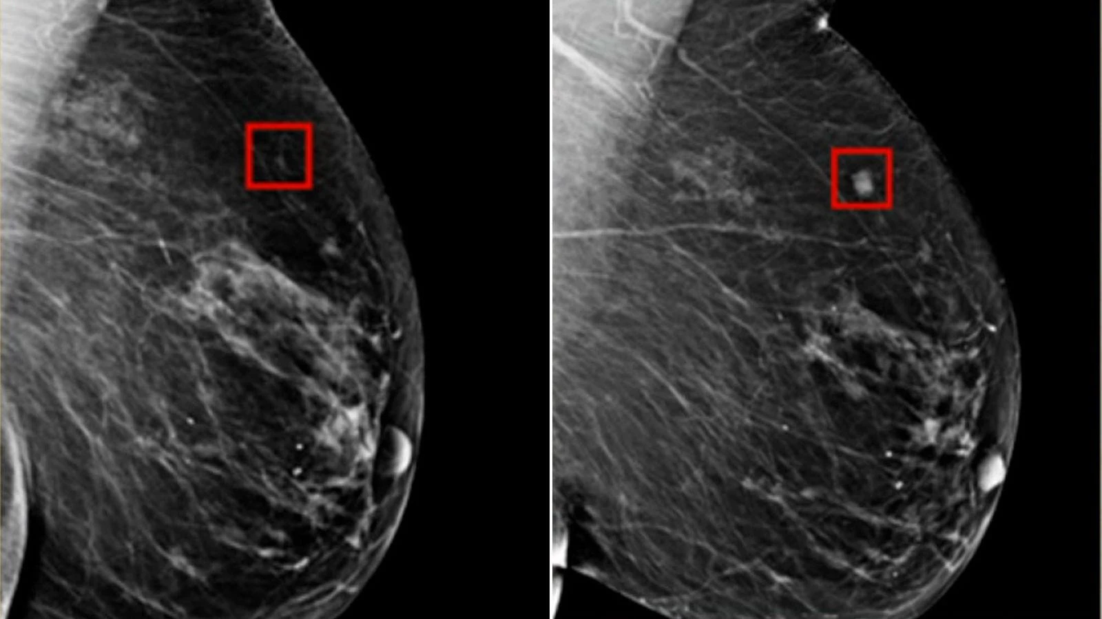
AI Revolution in Breast Cancer Diagnosis: How Cutting-Edge Technology is Enhancing Accuracy and Treatment Decisions

By Malikeh Sharifi
Did you know breast cancer is the second most common cancer worldwide and the number one cancer in women? Global statistic from 2022 show 2.3 million new cases reported, with 670,000 deaths caused by breast cancer. While the alarming statistics surrounding breast cancer highlight the urgent need for improved detection and treatment strategies, recent advancements in artificial intelligence are paving the way for more accurate diagnoses, partially in cases of ductal carcinoma in situ (DCIS). MIT and ETH Zurich researchers have developed an innovative AI model aimed at enhancing the diagnosis of ductal carcinoma in situ (DCIS). DCIS is a condition where abnormal cells are found in the lining of a breast duct, and while it is not yet invasive, it has the potential to become so if left untreated. Accurately diagnosing the stage of DCIS is crucial because it helps in determining the appropriate treatment and can prevent unnecessary interventions, which can be invasive and costly.

The traditional process of diagnosing DCIS relies heavily on the visual assessment of tissue samples by pathologists, who look for specific cellular patterns that indicate the presence and stage of cancer. This manual process can be subjective and prone to variability between different pathologists, leading to potential discrepancies in diagnosis and treatment plans. The new AI model aims to standardize and improve this process by analyzing the spatial organization and states of cells within tissue samples, offering a more objective and consistent diagnostic tool.

The AI model uses chromatin staining, a cost-effective imaging technique, to capture detailed images of the tissue samples. Chromatin, a complex of DNA and proteins found in the nucleus of cells, plays a key role in regulating gene expression, and its organization can provide important clues about the state of the cells. The AI then applies machine learning algorithms to these images to identify patterns that correspond to different stages of DCIS. This method allows for a more nuanced understanding of the disease, potentially leading to more accurate diagnoses.
One of the key benefits of this AI model is its scalability. Unlike more complex and expensive imaging techniques, chromatin staining is relatively simple and affordable, making it accessible for widespread use in clinical settings. This opens up the possibility of implementing the model in various healthcare environments, including those with limited resources, where traditional diagnostic tools may not be as readily available. Additionally, the approach could be adapted to diagnose other types of cancer or diseases, broadening its potential impact on healthcare.

The development of this AI model represents a significant step forward in the field of medical diagnostics. By providing a more accurate and reliable method for diagnosing DCIS, it has the potential to reduce overtreatment and improve patient outcomes. As research continues, the model may be further refined and integrated into clinical practice, offering a powerful tool for pathologists and helping to ensure that patients receive the most appropriate care for their condition.


interesting article!
Your article about the revolution of AI in breast cancer diagnosis was interesting. Knowing about the wonders AI technology has been doing in the past years is very important, especially when it comes to the medical field.